Can melanoma be only 2 mm across?
Plain-English answer
Yes. A melanoma can be only a few millimetres across. It may be flat, subtle and easy to overlook.
The familiar ABCDE warning signs are useful, although no single feature can rule melanoma in or out. The “D” refers to diameter. Melanoma can be smaller than 6 mm, and Cancer Council includes smaller spots that are growing as a possible warning sign.[1]
This case involved a melanoma in situ that was only about 2 mm across. The patient had not seen it. We found it because a conversation about his history led to a full skin examination.
A tiny spot found during a full skin check
A patient in his 70s recently mentioned that he had spent decades working outdoors. That can add up to a great deal of ultraviolet exposure, so I suggested that he return for a dedicated skin check.
One small pigmented spot caught my attention during the examination. It measured approximately 2 mm across. The patient had not noticed it and had no concerns about that area.
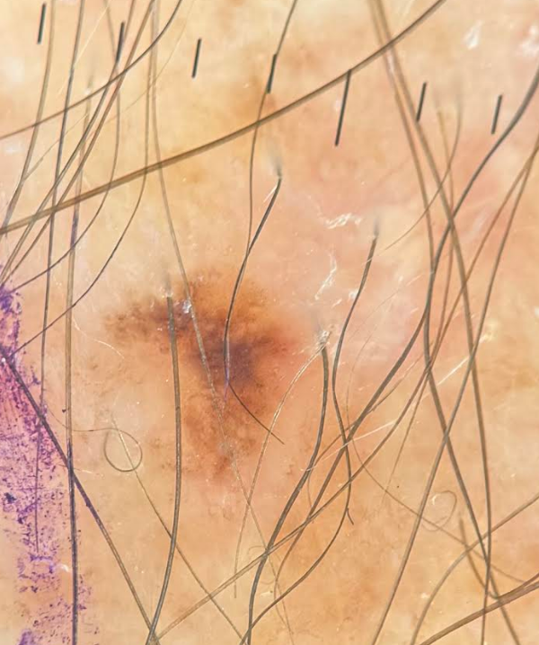
Dermoscopy image of the lesion described in this article. The image is highly magnified. The spot measured only approximately 2 mm across. Published with the patient’s explicit written consent.
Dermoscopy showed subtle asymmetry. The pigment and internal structure were unevenly distributed, with a darker structureless area sitting off-centre. These features raised enough concern to remove the spot and send it to a pathologist.
Histopathology confirmed melanoma in situ. The required excision was completed in general practice within three weeks of the first skin assessment.
This pathway suited this particular lesion and patient. Selected melanoma in situ and early-stage melanomas can be excised in primary care when the location is suitable and the doctor has the necessary training. Other melanomas need specialist or multidisciplinary care.[2]
What does melanoma in situ mean?
Melanoma in situ is also called stage 0 melanoma. The abnormal melanocytes remain within the epidermis, the top layer of the skin. They have not invaded the deeper dermis.[3]
At this stage, the melanoma has no route to spread elsewhere in the body. Complete excision is still required because the cells may eventually grow deeper if left untreated.
Why size can be misleading
Many people have heard that a mole larger than 6 mm deserves attention. Over time, that message has sometimes been interpreted to mean that smaller spots are safe.
Diameter is one clue among several. When I assess a spot, I also consider:
- whether it is new or changing
- asymmetry of its shape or internal structure
- an irregular border
- uneven colour or multiple colours
- whether it looks different from the patient’s other spots, sometimes called the “ugly duckling” sign
- symptoms such as bleeding, crusting or persistent itching
- the person’s age, skin type, sun exposure, number of moles, immune status and personal or family history of skin cancer.
Some melanomas fall outside the usual ABCD pattern. They may be pink or skin-coloured. Nodular melanomas are often assessed using the EFG clues: elevated, firm and growing.[1]
Most tiny brown spots are benign. Size simply needs to be considered alongside the rest of the clinical picture.
What dermoscopy adds
A dermatoscope is a handheld instrument that magnifies a lesion and reduces glare from the skin surface. It lets a trained clinician examine colours and structures that can be difficult to see with the naked eye.
The spot looks much larger in this photograph because it has been magnified. I focused on the uneven distribution of its colour and internal structure.
Research suggests that trained clinicians using dermoscopy assess possible melanoma more accurately than they do with visual inspection alone.[4] Dermoscopy guides the decision to leave a lesion alone, photograph and monitor it, or perform a biopsy. Pathology provides the diagnosis.
You can read more about the process in How doctors assess moles, spots and freckles.
Why I suggested a full skin check
This spot was an incidental finding. The patient’s long history of outdoor work came up during an ordinary consultation and led to the skin check.
Years spent outside can involve substantial UV exposure. I considered that history alongside his age and the appearance of his skin. An individual melanoma cannot be attributed to a particular job or period of sun exposure. The history still helped guide his risk assessment.
Other recognised risk factors include fair or freckled skin, significant UV exposure earlier in life, numerous or atypical moles, previous skin cancer, a strong family history and immunosuppression.[1]
This is one of the benefits of continuity in general practice. A passing detail can lead to a useful preventive-health assessment that otherwise may never have happened.
Does everyone need a routine full skin check?
No. The RACGP recommends matching the type and frequency of skin checks to each person’s risk.
Regular clinician skin checks are generally not recommended for people at average or below-average risk. Opportunistic skin examination is conditionally recommended for people at above-average risk. People at high risk should usually be checked at least annually, while those at very high risk may need six-monthly full skin examinations supported by total-body photography and dermoscopy.[5]
A GP can help work out your individual risk and whether you would benefit from a full skin examination.
What you can do between appointments
Much of the skin is difficult to inspect yourself. The back, scalp, behind the ears and backs of the legs are common blind spots. A mirror, photographs or help from a partner can make changes easier to identify.
Become familiar with your usual pattern of spots. Arrange a medical assessment if you notice one that is:
- new or clearly different from the others
- changing in size, shape or colour
- growing, even if it remains smaller than 6 mm
- persistently bleeding, crusting, itching or failing to heal
- concerning to you for any other reason.
Comparing your spot with photographs online is unlikely to give you a reliable answer. Melanomas vary considerably, and harmless lesions can sometimes look alarming.
The lesson from this case
This melanoma was approximately 2 mm across. The patient had not noticed it, and its appearance was subtle.
Its size formed only one part of the assessment. The patient’s background, a complete examination and the structures seen under dermoscopy led to the decision to remove it. Pathology then provided the answer.
Early melanoma can be subtle. In this case, the starting point was a conversation about years spent working outside.
References
- Cancer Council Australia. Melanoma signs, symptoms and risks. Updated March 2025.
- Cancer Council Australia. Optimal care pathway for people with melanoma, second edition. 2021.
- Cancer Council Australia. Diagnosing melanoma. Updated March 2025.
- Dinnes J, Deeks JJ, Chuchu N, et al. Dermoscopy, with and without visual inspection, for diagnosing melanoma in adults. Cochrane Database Syst Rev. 2018;12:CD011902.
- Royal Australian College of General Practitioners. Preventive activities in general practice: skin cancer. Updated 28 June 2024.
This article provides general information only and is not a substitute for individual medical advice.